Cancer is a leading cause of death worldwide, and early detection is key to successful treatment. However, detecting cancer can be challenging, especially in early stages when the cells are less abnormal. In an effort to improve cancer detection, Google and the Department of Defense (DoD) have teamed up to develop an AI-powered microscope.
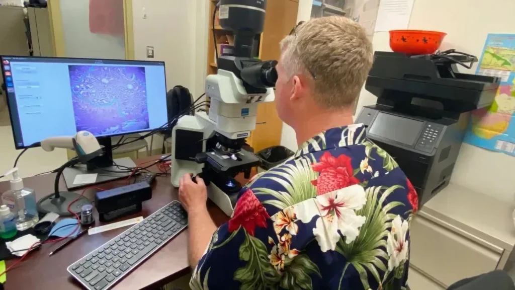
The microscope, called the Augmented Reality Microscope (ARM), uses artificial intelligence to identify cancerous cells in tissue samples.
The ARM works by using computer vision algorithms to analyze the tissue samples. The algorithms have been trained on a massive dataset of cancer images, and they are able to identify cancerous cells with high accuracy.

Source: U.S. Department of Defense
When a pathologist uses the ARM to examine a tissue sample, the AI algorithms overlay a heatmap onto the image. The heatmap shows the pathologist where the AI is most likely to have identified cancerous cells. The pathologist can then use this information to focus their examination on the most suspicious areas of the sample.
The ARM is still under development, but it has the potential to revolutionize cancer detection. By helping pathologists to identify cancerous cells more quickly and accurately, the ARM could help to save lives.
In this blog post, we will take a closer look at the ARM and its potential impact on cancer detection. We will also discuss the challenges that lie ahead in bringing the ARM to market.
Contents
AI-Powered Microscope: Importance Of Microscopy
Microscopy is important in many different fields, including biology, medicine, materials science, and engineering. In biology, microscopy is used to study cells, tissues, and organs. In medicine, microscopy is used to diagnose diseases, monitor the progression of treatments, and conduct research. In materials science and engineering, microscopy is used to study the structure and properties of materials.
Here are some specific examples of the importance of microscopy:
- Biology: Microscopy is essential for studying cells, which are the basic building blocks of life. Microscopes allow us to see the structure and function of cells, as well as the interactions between cells. This knowledge is essential for understanding biology and developing new treatments for diseases.
- Medicine: Microscopy is used to diagnose a wide range of diseases, including cancer, infections, and autoimmune diseases. Microscopes are also used to monitor the progression of treatments and to conduct research on new treatments.
- Materials science and engineering: Microscopy is used to study the structure and properties of materials. This information is used to develop new materials with improved properties, such as strength, durability, and lightness.
Microscopy is a powerful tool that has revolutionized our understanding of the world around us. It continues to be used to make new discoveries in science and medicine, and to develop new products and technologies.
In addition to the above, microscopy is also important in education and research. Microscopes allow students to see and understand the world around them in a more detailed way. They are also essential for conducting research in a wide range of fields, including biology, chemistry, physics, and materials science.
Objectives of Google and the Department of Defense (DoD) in building an AI-powered Microscope
The AI-powered microscope also has the potential to improve cancer diagnosis and treatment in other ways. For example, it could be used to develop new biomarkers for cancer detection, to assess the aggressiveness of tumors, and to predict how patients will respond to different treatments.
Main Highlights:
- Improve the accuracy and speed of cancer detection. Cancer is a leading cause of death worldwide, and early detection is key to successful treatment. The AI-powered microscope can help pathologists to identify cancerous cells more quickly and accurately, which could lead to earlier diagnoses and better patient outcomes.
- Reduce the workload on pathologists. Pathologists are in high demand, and they are often overworked. The AI-powered microscope can help to reduce the workload on pathologists by automating some of the tasks involved in cancer detection. This could free up pathologists to focus on more complex cases and to conduct research.
- Make cancer detection more accessible. The AI-powered microscope is relatively inexpensive to produce, and it can be used in a variety of settings, including hospitals, clinics, and even remote locations. This could make cancer detection more accessible to people who live in underserved areas or who have difficulty traveling to see a pathologist.
Overall, the AI-powered microscope has the potential to revolutionize cancer detection and treatment. It is a promising new technology that is being developed by Google and the DoD in collaboration with pathologists and other experts.
Google’s AI-Powered Microscope Features & Applications
- Automated image analysis: AI-powered microscopes can automatically analyze images of cells and tissues, identifying features that would be difficult or time-consuming for human pathologists to identify manually. This can help to improve the accuracy and speed of diagnosis.
- Real-time feedback: AI-powered microscopes can provide real-time feedback to pathologists as they are examining slides. This can help pathologists to identify suspicious areas more quickly and to focus their attention on the most important parts of the slide.
- Improved image quality: AI-powered microscopes can improve the quality of microscope images by reducing noise and blurring. This can make it easier for pathologists to see important features of cells and tissues.
- Quantitative analysis: AI-powered microscopes can perform quantitative analysis on images of cells and tissues. This can help pathologists to measure features such as cell size, shape, and texture. This information can be used to diagnose diseases and to monitor the progression of treatments.
Here are some potential applications of the AI-powered microscope:
- Cancer detection: AI-powered microscopes can be used to automatically detect cancerous cells in tissue samples. This can help pathologists to diagnose cancer more quickly and accurately.
- Infectious disease diagnosis: AI-powered microscopes can be used to diagnose infectious diseases by identifying pathogens in tissue samples or blood smears.
- Drug discovery: AI-powered microscopes can be used to screen drug candidates for their ability to kill cancer cells or to inhibit the growth of bacteria.
- Research: AI-powered microscopes can be used to study the biology of cells and tissues in a more detailed way. This could lead to new insights into the causes and treatments of diseases.
Ethical Considerations:
- Accuracy and reliability: AI-powered microscopes are still under development, and it is important to ensure that they are accurate and reliable before they are used in clinical settings. This means testing them on large datasets of known cases and ensuring that they can perform at a level that is comparable to human pathologists.
- Bias: AI-powered microscopes are trained on data that is collected from human pathologists. This data may contain biases that are reflected in the AI system. It is important to be aware of these biases and to take steps to mitigate them.
- Transparency: It is important to be transparent about how AI-powered microscopes work and how they are being used. This is important for both patients and healthcare professionals.
- Accountability: There should be clear lines of accountability for the use of AI-powered microscopes. This means that it should be clear who is responsible for the accuracy and reliability of the system, as well as for any decisions that are made based on the results of the system.
In addition to these general ethical considerations, there are also some specific concerns that have been raised about AI-powered microscopes. For example, some people have expressed concern that these systems could be used to develop new forms of surveillance or to discriminate against certain groups of people.
Conclusion
The AI-powered microscope that is being developed by Google and the Department of Defense has the potential to revolutionize cancer detection. By helping pathologists to identify cancerous cells more quickly and accurately, the AI-powered microscope could lead to earlier diagnoses and better patient outcomes.
The AI-powered microscope also has the potential to make cancer detection more accessible and affordable. The system is relatively inexpensive to produce, and it can be used in a variety of settings, including hospitals, clinics, and even remote locations. This could make cancer detection more accessible to people who live in underserved areas or who have difficulty traveling to see a pathologist.
While the AI-powered microscope is still under development, it has the potential to make a significant impact on the fight against cancer. By improving the accuracy, speed, and accessibility of cancer detection, the AI-powered microscope could help to save lives.
In addition to its potential impact on cancer detection, the AI-powered microscope also has the potential to improve cancer diagnosis and treatment in other ways. For example, it could be used to develop new biomarkers for cancer detection, to assess the aggressiveness of tumors, and to predict how patients will respond to different treatments.
Must Read:
Google Pixel Watch 2: Release Date, Specs, and Price
Overall, the AI-powered microscope is a promising new technology with the potential to make a significant impact on the fight against cancer. It is a collaboration between Google and the DoD in partnership with pathologists and other experts.


